Improving the Speed of MRI

UB researcher receives award from SUNY Technology Accelerator Fund to improve magnetic resonance imaging.
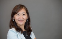
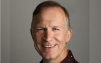
Leslie Ying is improving magnetic resonance imaging. For this research, she will receive $50,000 each from the statewide SUNY Technology Accelerator Fund (TAF), which cultivates innovation by speeding the commercialization of high-impact SUNY inventions.
The project is one of five selected from across the SUNY system and announced last month by Gov. Andrew M. Cuomo, SUNY and the SUNY Research Foundation.
A quicker, less redundant MRI scan
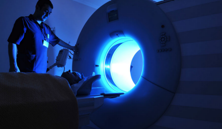
Medical imaging is the magic process of looking inside the body without making a cut. But it’s only as good as the picture you get — the better the picture, the better the doctor can determine what’s wrong with the patient.
Magnetic resonance imaging (MRI), one of the most widely used forms of medical imaging, draws on physics, math and engineering, says Leslie Ying, associate professor of biomedical engineering and electrical engineering.
“My role is computation,” she says. “I develop innovative algorithms for MRI to make the image look better.”
Better images are one thing, but Ying’s goal is to make it happen more quickly. “The issue with images and speed is that the patient has to stay in the scanner — motionless — for a period of time,” she says. Sometimes, that means patients are asked to hold their breath. Other times, doctors may want to look at an organ that can’t be stilled, such as a beating heart.
Shorter times also can mean cost savings, Ying says. So her challenge, she says, is: “Can I significantly reduce this period of time?”
Ying’s method uses a complicated algorithm to generate an image from only a small portion of the data that is commonly collected. That leaves another aspect of the challenge: Can Ying’s method produce the same image quality as a longer scan?
A lot of data in an image — whether an MRI scan or a vacation photo — is redundant, Ying says. In digital photography, people often compress image data with a file format, such as JPEG, that makes a file size smaller with minimal loss of quality.
Ying’s method is analogous to that, except that it predicts image redundancies in a process she calls compressed-sensing. “The idea is that we don’t acquire all the information in the first place,” she says. “We anticipate what compression will do and we only look for those points.”
To do that without guessing, Ying’s method starts with a model. She then needs only a few data points to complete the information. “Our technique improves the speed of scanning,” Ying says, noting her work has validated the algorithms used in her methods and has demonstrated proof of concept.
Now, with the TAF funding, Ying and her team will test her method in an MRI scanner — “to see really how long it takes. A few seconds? A minute? We need to be able to demonstrate to vendors what we can offer.”
Ying will work with colleagues at GE Healthcare in Wisconsin — she was formerly at the University of Wisconsin, Milwaukee — to run the tests.
“I have always been fascinated by what medical imaging can do. I want to contribute directly to that,” Ying says. “If an algorithm is too complicated, it’s not going to work. I try to find the balance and fill the gap between academic work (mathematical theory) and industrial practice (medical imaging).”
Published May 20, 2014 This content is archived.